
 PDF Download
 PubReader
 Export Citation
 Email the Author
 Share Facebook
 Share Twitter
 Cited by 0 Article
 Metrics (View:2,476)






1. INTRODUCTION
In situ chemical sensing remains an important challenge in healthcare and environmental monitoring due to the need for continuous, selective, and power-efficient operation [1–3]. Compared with purely physical transducers, emerging chemical and biochemical sensors present distinct challenges because their operation is inherently more complex and typically involves additional transduction steps. Conventional chemical and biochemical sensing architectures typically rely on separate recognition and transduction elements, increasing system complexity [3]. Chemical recognition elements often dominate device complexity because they must translate molecular interactions into measurable physical signals. Despite substantial advances in chemical/biochemical sensing technologies, receptors remain vulnerable to coexisting interferents and target species and are often consumable or non-renewable [4].
Recently, hydrogels have attracted considerable interest as receptor elements [5,6]. Hydrogels are water-swollen polymer networks that can be engineered with functional groups responsive to specific environmental stimuli. Such hydrogels exhibit reversible swelling or shrinkage in response to chemical stimuli, including pH and ionic composition [6–8]. Such materials are appealing as receptors because they operate passively without an onboard power source, can be miniaturized, and are amenable to integration with relatively simple readout architectures. When combined with advancing microelectromechanical systems (MEMS) technology, hydrogels have enabled a variety of novel chemical/biochemical sensing modalities [9–11]. Prior studies have demonstrated MEMS-based sensing platforms in which hydrogel swelling modulates electrical parameters such as capacitance [9]. However, several bottlenecks limit broader deployment of such devices. Fabrication of hermetically sealed MEMS transducers generally requires cleanroom processing, and the achievable wireless range becomes inadequate for deeply implanted locations (e.g., bladder, gastrointestinal tract, or arteries) because the relatively high operating frequencies (~MHz) experience substantial attenuation in tissue [12].
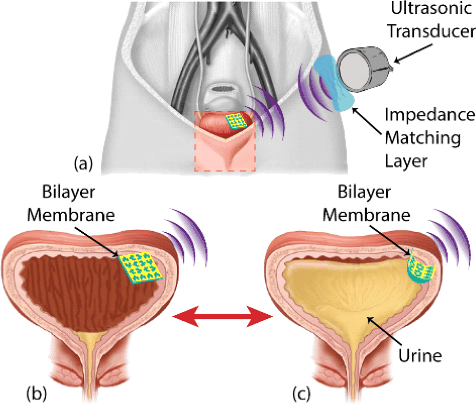
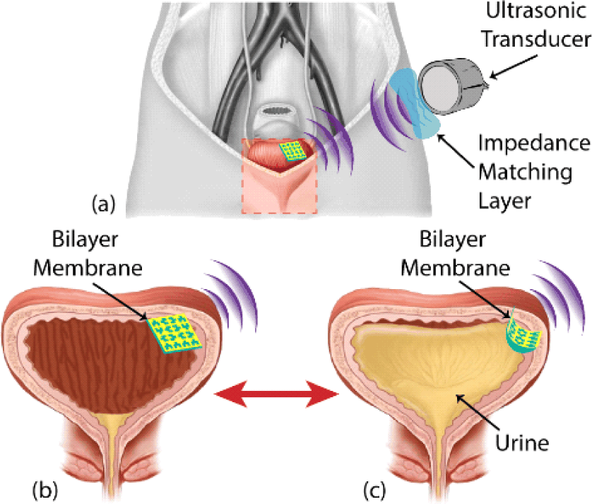
Here, we present a hydrogel–fractal bilayer piezoelectric thin film that enables physiological sensing through deformation-induced ultrasonic modulation. Schematics of the bilayer device to monitor urinary pH for early detection of urinary tract infection (UTI) are given in Fig. 1. Normal urine is typically slightly acidic, with pH values between 4.5 and 8 [13]. In the presence of urea-splitting organisms such as Ureaplasma urealyticum, Proteus, or Klebsiella, the pH level in the bladder can shift toward alkaline values [14]. Ambulatory bladder pH monitoring could therefore enable early detection and prevention of UTIs. The detection mechanism (Fig. 1(b)–Fig. 1(c)). The thin film can be rolled into a cylindrical form and delivered into the bladder through the urethra. Once deployed, filling the bladder with urine causes the pH-sensitive hydrogel to swell or shrink; elevated pH induces curvature in the bilayer, which, in turn, alters the ultrasonic response, including amplitude, frequency, and focal characteristics. In such a transduction scheme, the pore architecture of the fractal membrane is critical: (1) it enhances acoustic power output by increasing plate mobility [15]; (2) it broadens bandwidth by supporting additional vibrational modes [16]; and (3) it increases the attainable curvature by providing void volume for hydrogel expansion. Encoding chemical information into the ultrasonic response provides an alternative sensing pathway that avoids several limitations of conventional biochemical sensors. Ultrasonic interrogation enables wireless readout from deeply implanted sites while maintaining a simple external receiver, and the device can be fabricated without reliance on conventional cleanroom-based MEMS processes.
2. METHODS
A fractal is a set of patterns that are self-similar across multiple length scales, a phenomenon that is frequently observed in nature [1]. Numerous fractal patterns have been derived from mathematical models, including Hilbert, Sierpiński, Peano, Moore, von Koch, Vicsek, and Greek cross curves [2]. These patterns have been applied to enhance the stretchability of flexible electronic structures, as demonstrated by Fan et al. [3]. In a related study, Putra et al. showed that introducing pore architectures into flexible panels improves plate mobility [4].
Motivated by these findings, we implemented fractal designs in piezoelectric transducers to increase power output by enhancing plate mobility. Our group previously reported that fractal ultrasonic transducers, such as polyvinylidene difluoride (PVDF) transducers patterned with fractal geometries, generate higher output power and exhibit increased bandwidth due to the additional vibration modes enabled by the fractal design. Consequently, both frequency and acoustic intensity responses were improved [5]. While our prior work focused on fractal PVDF membranes as enhanced electromechanical ultrasonic transducers, the present study extends this concept by integrating the fractal membrane with a pH-responsive hydrogel to form a functional bilayer for biochemical sensing. In this architecture, hydrogel swelling or shrinkage induces curvature in the bilayer, thereby modulating the vibration behavior and ultrasonic output of the fractal piezoelectric membrane. This bilayer integration establishes a chemo-mechanical to mechano-electrical transduction pathway, transforming the fractal transducer from a passive electromechanical device into an active, stimulus-responsive platform for battery-less biochemical sensing.
As a proof of concept, we employed a modified Hilbert space-filling curve on the hydrogel–fractal bilayer transducer. The Hilbert curve is a continuous fractal space-filling curve [6], as illustrated in Fig. 2(a).
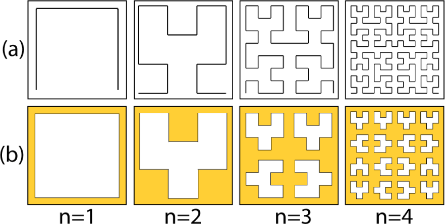
Because conventional Hilbert curves are open structures, we slightly modified them to form closed shapes while preserving the periodicity of the original Hilbert pattern. Modified Hilbert space-filling curves with closed geometries were chosen to optimize both electromechanical performance and bilayer integration. The high pore density enhances plate mobility and supports multiple localized vibration modes, resulting in increased bandwidth, while the closed-loop architecture preserves structural and electrical continuity of the piezoelectric membrane. In addition, the internal void volume accommodates hydrogel swelling and shrinkage without inducing mechanical delamination, making these patterns well suited for hydrogel–fractal bilayer transducers. Fig. 2(b) shows the modified version of Hilbert curves for closed shapes in iteration orders (n) of 1 through 4. The iteration order indicates the number of intervals overlapping with a segment of the curve. The fractal portion of the hydrogel–fractal bilayer transducer was designed using these space-filling patterns.
The bilayer device includes a hydrogel layer as its second component. Hydrogels consist of crosslinked, three-dimensional networks of long polymer chains that are inherently hydrophilic. This hydrophilic nature can be exploited by incorporating targeted chemical moieties into the polymer network, rendering the hydrogel sensitive to environmental parameters such as pH, specific ions, glucose, or antigens [7–9].
Hydrogels are attractive as receptor materials because they are biocompatible, reusable, have a small form factor, and are chemically activated, thereby eliminating the need for onboard power. pH-responsive hydrogels have previously been employed in implantable sensors [10–14]. The hydrogel used in this work for laminating the fractal piezoelectric transducer is a poly(methacrylic acid)-based hydrogel that undergoes reversible swelling and shrinking when exposed to different pH environments. The detailed formulation of this hydrogel is described in the fabrication section of this manuscript.
In acidic media (pH<7.0), the hydrogel film shrinks, whereas in basic media (pH>7.0), it absorbs more water from the environment and swells, thereby generating the desired chemo-mechanical response. During this volume phase transition, the attached flexible piezoelectric film deforms to conform to the hydrogel, causing the curvature of the piezoelectric layer to increase or decrease relative to the neutral (flat) state. This curvature change modifies the acoustic intensity response of the bilayer transducer. The resulting change in ultrasonic response can be interrogated using an external ultrasonic receiver, and the measured output can be correlated to the pH of the surrounding medium.
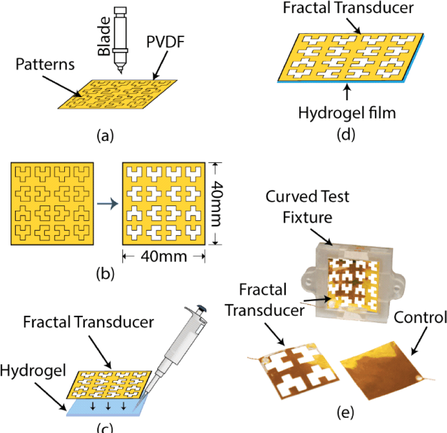
Bilayer transducers were fabricated by patterning a perforated piezoelectric membrane followed by hydrogel integration. The overall process is summarized in Fig. 3.
We first selected the fractal pattern to be implemented. In this proof-of-concept study, we chose modified Hilbert space-filling curves because they are among the simplest fractal curves that can be readily adapted to form closed shapes, as shown in Fig. 2(b). The closed geometry allows the creation of perforations on the transducer surface. Third- and fourth-order Hilbert space-filling curves were implemented on the piezoelectric transducers, as these higher orders have been shown to generate larger vibration amplitudes than first- and second-order patterns [5].
The Hilbert patterns were modified to form closed shapes using CAD software. A tungsten-blade cutter/ plotter (CAMEO3, Silhouette Inc.) was then used to cut these patterns into a 50 μm-thick PVDF piezoelectric polymer membrane (Fig. 3(a), Fig. 3(b)). After forming the perforated structures, electrical connections were established using two 30-gauge insulated copper wires by silver epoxy (8331, MG Chemicals).
The pH-responsive hydrogel formulation was adapted from previously reported methods [15] and was carried out in two parts (solutions A and B). The base solution was prepared by dissolving acrylamide (334.5 mg, AAm, Sigma-Aldrich), methacrylic acid (100.8 μL, mAA, Sigma-Aldrich), TEMED (100 μL, Sigma-Aldrich), and N,N′-methylenebisacrylamide (16.35 mg, Sigma-Aldrich) in DI water (1.2 mL). The mixture was stirred until all components were fully dissolved. The curing solution was prepared separately by dissolving ammonium persulfate in DI water (80 mg/mL).
The pH-responsive hydrogel was prepared by mixing base and curing solutions at a ratio of 5.9:1. Immediately before lamination, the mixture was combined at this ratio and mixed vigorously in a beaker using a vortex mixer to ensure uniform dispersion, then poured into a substrate, followed by placing the fractal PVDF transducer immediately, as gelation begins rapidly (~60 s). Due to gravity, a uniform ~1 mm-thick hydrogel layer formed and adhered to the fractal transducer after polymerization (Fig. 3(c), Fig. 3(d)), yielding a hydrogel–fractal bilayer transducer.
Three types of hydrogel–fractal bilayer transducers were fabricated: devices with third-order Hilbert patterns, devices with fourth-order Hilbert patterns, and non-fractal devices (no perforations) as control samples. The fabricated samples are shown in Fig. 3(e), which also includes the experimental apparatus and the fourth-order bilayer transducer used for subsequent characterization.
The prototype hydrogel-fractal piezoelectric bilayer transducer was characterized for wireless biochemical sensing. The experimental setup is illustrated in Fig. 4. All characterization experiments were conducted in the water tank (Precision Acoustics UK) (Figure 4(b)). Note that water media have acoustic properties similar to those of soft tissue [16]. We started by investigating the chemomechanical response (curvature change due to the pH change of the medium) of the bilayer transducer. Then we also studied the natural frequency of different bilayer transducers and characterized the ultrasonic response from the bilayer transducers under different pH environments. For characterizing the ultrasonic response, a custom frame was manufactured to hold the protype to direct its output towards the ultrasonic receiver (Fig. 3(e)). The frame could hold the prototypes in line with the receiver at specific curvature angles (θ). The curvature angle was created due to pH change of the medium (Fig. 4(a)) and was determined by θ=S/r, where S is the arc length; r is the radius, and θ is the central angle of curvature. For our experiments, arc length, S=40 mm, and central angle, θ=0°, 30°, 60°, and 120°.
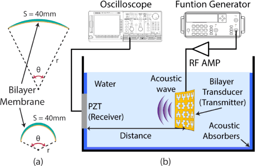
The hydrogel fractal piezoelectric bilayer transducer was excited by a pulsed electric signal (V=0.225 V, frequency=5 kHz, pulse width=0.25 ms) using a function generator (4065, B&K Precision) to generate ultrasonic waves. The generated ultrasonic wave travels through the water in the tank and reaches another ultrasonic transducer (PZT5H, Piezo Inc.) affixed to the side of the tank as a receiver. The received ultrasonic intensity on the receiver transducer is a function of distance and curvature angle. The curvature angle also depends on the specific pH level of the medium (water in this case). The distance between the transmitter and receiver transducer was also varied between 20 mm and 90 mm. It is important to note that, to simplify the experiment, the bilayer transducer was excited electrically by an external source, whereas in real cases an ultrasonic transducer would transmit ultrasound to the bilayer transducer and detect the reflected ultrasonic waves.
3. RESULTS AND DISCUSSION
The hydrogel is responsive to the pH of the surrounding medium. Changes in pH alter the swelling state of the hydrogel and thus change the curvature of the bilayer. We evaluated this response in different pH solutions (pH 4–12) and measured the corresponding curvature angles.
Fig. 5 presents time-resolved optical images of the bilayer transducers under each pH condition. As evident from the images, exposure to different pH solutions caused the bilayer to curl, and the resulting curvature angle could be visually observed and quantified. The hydrogel shrank in more acidic media (lower pH) and swelled in more basic media (higher pH) in a reversible manner. Consequently, larger curvature angles were obtained at higher pH values.
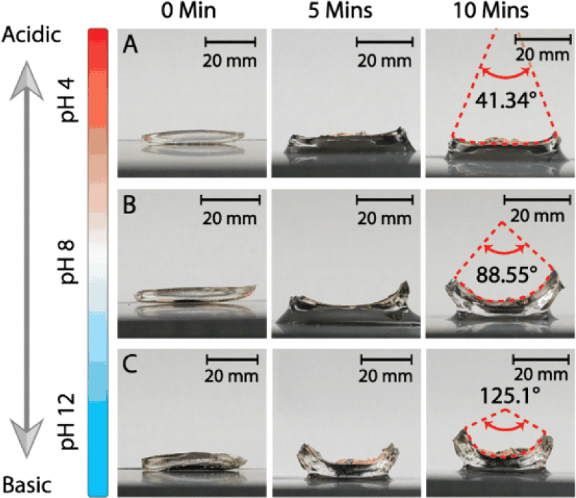
The curvature angles measured at 10 min (approximately the time at which swelling/shrinking reached steady state) were 41.3°, 88.6°, and 125.1° for pH 4, 8, and 12, respectively. From these data, the responsivity of the bilayer transducer was calculated to be 10.47°/pH.
Two sets of experiments were conducted to characterize the mechanoelectrical response. First, we measured the frequency response of the bilayer transducer (Fig. 6).
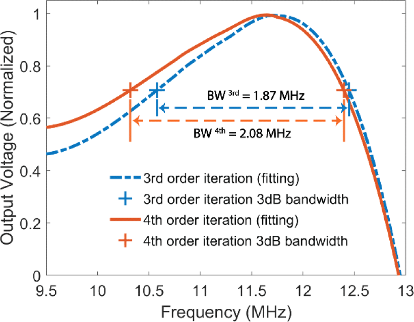
The resonance frequencies with Hilbert iteration orders n=3 and 4 were both approximately 11.6 MHz. The −3 dB bandwidths (where the received ultrasonic intensity was reduced by half, indicated by ‘+’) were 1.87 MHz and 2.08 MHz for the third- and fourth-order samples, respectively. The results indicate that higher Hilbert iteration order correlates with increased bandwidth, consistent with the enhanced plate mobility near the edges of the pore architecture [4,5]. As the fractal order increases, the surface perforation density increases, enabling the transducer to vibrate in multiple directions and to support more localized vibration modes with different amplitudes. These additional modes lead to higher overall vibration and a broader bandwidth compared to the non-perforated (non-fractal) design.
We evaluated the ultrasonic output response using the receiver configuration shown in Fig. 4. The curved hydrogel–fractal piezoelectric bilayer transducer is expected to generate distinct ultrasonic responses for different curvature angles corresponding to different pH levels. We quantified the responses in terms of received voltage per unit area of the transmitter (mV/cmM2) for multiple pH levels (curvature angles) and for different Hilbert iteration orders (n=3,4), as well as for the non-fractal control sample.
In these experiments, the curvature angles were set to 0°, 30°, 60°, and 120° using the custom fixture shown in Fig. 3(e). These angles were chosen both for experimental convenience and to span the range observed in the chemomechanical experiments (41.3°–125.1° for pH 4.0–12.0).
The bilayer sample was vertically translated to adjust the transmitter–receiver separation from 20 mm to 90 mm, with output signals recorded at 3-mm intervals. The corresponding time-of-flight delays were between 14 μs and 62 μs, assuming an acoustic velocity of 1,496 m/s in water at 25°C. To capture the distance-dependent ultrasonic response, output voltages were averaged across measurements at each separation distance.
Fig. 7 shows the logarithmic-scale output voltage as a function of curvature angle. As the Hilbert iteration order increased, the ultrasound response became more linear with respect to curvature angle, indicating improved linear responsivity at higher fractal orders. In contrast, the control sample (no fractal pattern) exhibited an exponential decrease in voltage with increasing curvature angle.
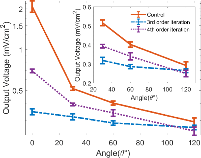
We calculated the sensitivity and linearity for each device. The third-order bilayer transducer exhibited improved linearity compared with the control, with a slope of 0.0006 mV/cm2/degree and R2=0.90. Bilayer transducer with fourth-order patterning showed even higher linearity and sensitivity, with a slope of 0.0016 mV/cm2/degree and R2=0.99. Accordingly, the wireless voltage sensitivities were determined to be 0.006 mV/cm2/pH for the third-order device and 0.017 mV/cm2/pH for the fourth-order device. The observed curvature-dependent ultrasonic response can be attributed to deformation-induced changes in the vibration behavior and radiation characteristics of the bilayer transducer. Hydrogel swelling or shrinkage introduces curvature that alters the effective stiffness, vibration mode shape, and acoustic radiation pattern of the fractal membrane. Increased curvature reduces effective plate mobility and modifies acoustic impedance matching with the surrounding medium, leading to a decrease in forward-directed ultrasonic intensity. These effects collectively explain the systematic reduction in received ultrasonic amplitude with increasing curvature observed in Fig. 7.
4. CONCLUSION
The proposed architecture provides a compact, low-cost, battery-free platform capable of long-range wireless biochemical sensing. Unlike prior fractal piezoelectric transducers that focused solely on enhanced electromechanical performance, the hydrogel–fractal bilayer presented here enables direct biochemical sensing by coupling pH-induced chemomechanical deformation with curvature-dependent ultrasonic modulation. By exploiting the reversible swelling behavior of pH-responsive hydrogels as receptor elements and integrating them with fractal PVDF piezoelectric transducers, we demonstrated that the sensing range, bandwidth, and wireless interrogation capability can be significantly enhanced. The fractal design improved both the frequency response (increased bandwidth) and the linearity of the mechanoelectrical transduction.
By combining chemomechanical and mechanoelectrical mechanisms, the proposed sensing scheme enables wireless transmission of pH information from the medium to an external interrogation unit without any onboard power source. The presented system offers a simple, low-cost, battery-less, and long-range bio-chemomechanical sensing platform that has the potential to overcome many limitations associated with conventional chemical and biochemical sensing technologies. Future work will evaluate the hydrogel–fractal bilayer transducer under physiologically relevant conditions, including the effects of ionic strength, temperature-dependent hydrogel kinetics, and mechanical constraints imposed by surrounding tissues. Understanding these factors will be essential for optimizing device performance and enabling reliable in-vivo implementation.


